I think there’s something oddly beautiful about vintage anatomy illustrations, and judging from the demand for them, I know I’m not alone! There is a multitude of uses for the free old anatomy drawings we’ve collected for you. Use them as clip art in a school project, tattoo inspiration, or reference pictures for life drawing! Then there’s Halloween – perhaps you need downloadable skeleton images for decorations or printable Halloween party invitations.
Old medical illustrations also look amazing simply printed and hung as wall art (especially vintage medical charts like the one above), and we’ve made sure they are high-resolution so you get the best results! I’ve seen people use them as prints on t-shirts, mugs, and even as quirky wallpaper, so whether you’re into skulls, skeletons, muscle structures or something even more specific in the body, you’ll find it here in our full vintage anatomy illustration gallery. Black and white anatomy graphics, or full-color vintage prints – they’re all over at the gallery, and we’ll be adding even more vintage anatomy pictures in the near future.
But every picture has a story! So here are a few of the best anatomy and vintage medical illustrations, and the stories and people behind them. WARNING! The study of anatomy has a grisly past, so if murder and body snatching isn’t quite your thing, maybe you’d prefer to check out our extensive animal illustration gallery instead?
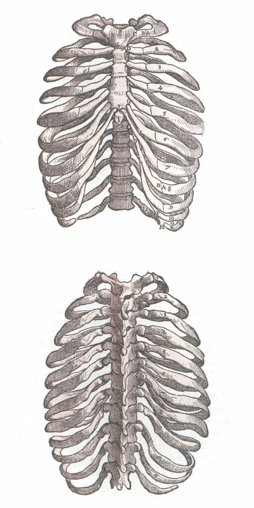
Old medical illustration of a black and white ribcage
I loooove myself some good ribs. And how great is this vintage ribcage medical illustration by Andreas Vesalius?! Here we have a clear image of a human ribcage from front and back, which means we can see the connection of the ribs with the sternum and the spine.
The ribs are numbered, which I’m assuming has a medical meaning. I like how the labeling of the ribs takes this from what would be an almost cartoonish depiction of the human ribcage to a proper scientific illustration.
Unlike today, where art and science are often seen as at odds, being a skilled illustrator came hand in hand with being a scientist. It is thought that Vesalius worked with artist Jan van Calcar to produce some of the illustrations for his work “Fabrica”.
Fun fact: Andreas Vesalius was a Flemish anatomist and is often referred to as the founder of the study of modern human anatomy. There’s even a crater on the moon named after him. We’ve got a whole heap more of his scientific drawings in our Andreas Vesalius gallery.
I think this would make a really funky greeting card to go with a gift for an orthopedic surgeon! Or printed and laser cut onto acrylic for a funky pair of halloween earrings.
Vintage skull illustrations from old books
There is something absolutely pleasing about a visual chart. They make me feel clever, informed, and organised, even when the subject matter is something as unsettling as an illustration of the skull!
The left image of a chart shows the labelled and numbered sections of the skull, and also of a complete male head, straight on and in profile. It appears to be comparing different types of tooth and jaw growth as well.
The chart on the right has illustrations of the skull in profile, facing us, and from the top down. It also has drawings of the skull broken into its separate pieces, with detailed labelling.
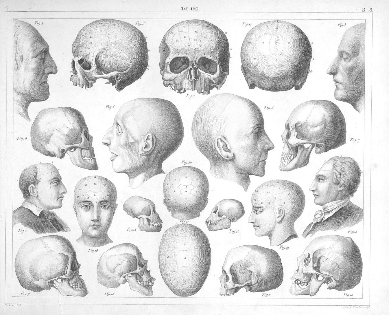
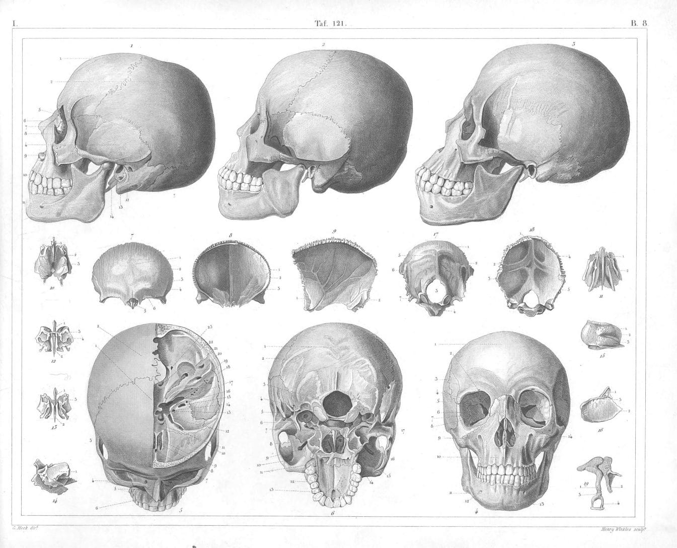
These two charts are by J.G. Heck and have been extracted from the book of antique illustrations Iconographic encyclopaedia of science, literature, and art. The antique book was a beast of a visual encyclopedia and included old illustrations of people, places, animals, and science.
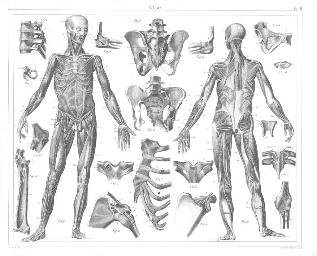
Full body old anatomy illustration showing muscles and organs
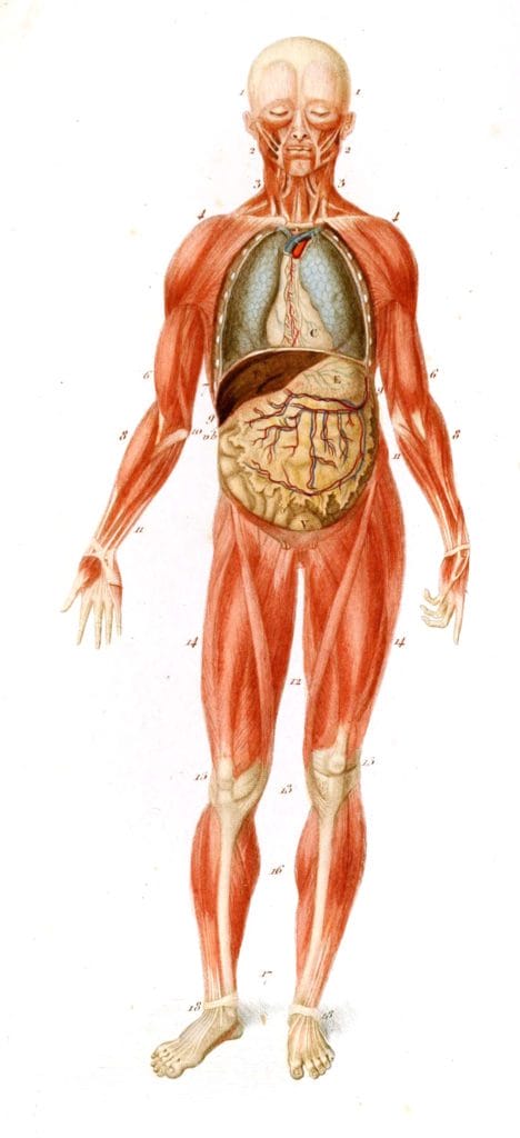
I’ve got you….under my skinnnnn…. This image is an antique medical diagram of the main muscles and organs in the body. I would assume this is a simplified view for clarity’s sake because the real focus here is to illustrate the organs, with the surrounding form of the body giving that context.
Again, I like that the diagram of this standing human body is labeled because it moves it clearly away from fine art and into the realm of science. It feels useful, informative, and reflective of the artist’s detailed knowledge of their subject.
I also, strangely, grotesquely maybe, like the color! The artist had used an unusually vibrant orange-red to color the muscles, and an antique beige and minty turquoise to tint the organs. The use of color makes this diagram even clearer to us. It’s likely that this would have been hand coloured with watercolour or ink.
This image is by Charles Dessalines D’Orbingy, who surprisingly was not an anatomist himself, but a zoologist and naturalist. His father was a physician though, so he may have had connections that led to him being commissioned for artistic work in the medical field.
Antique woodcut print of an early surgical table and tools
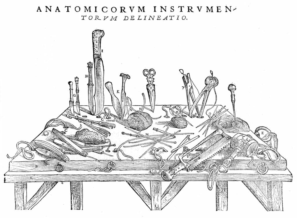
At first glance, this might seem like a messy bench in a garage or kitchen. But on closer inspection, you’ll see that this illustration is not for the faint-hearted.
Early anatomical research was not a pretty world to find yourself in, and these are the tools that doctors and scientists would have used to explore the human body. We can see surgical saws, scissors, knives in all shapes and sizes, a pounding mallet, twine, and a range of picks or needles. It’s doubly unsettling to think that what you see illustrated here would have been the same tools as used in surgery, with only crude natural remedies or alcohol used as anaesthetic.
This illustration of vintage surgical tools is another from Fabrica by Andrea Vesalius. Imagine this printed as gift wrap for your favorite surgeon!
Medical art? A vintage series of standing men with illustrated muscles and bones
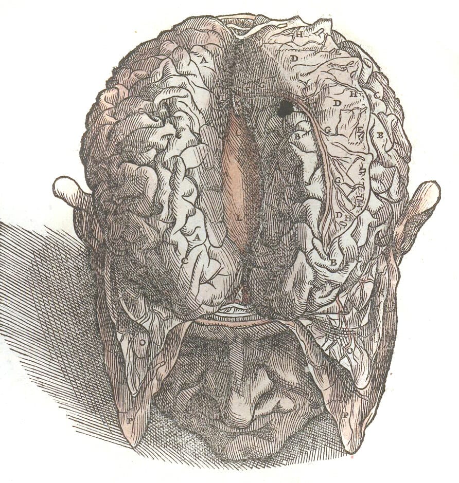
Speaking of gruesome, have a look at this! It’s another one by Andrea Vesalius, but this one have moved slightly away from what would be classed as medical illustration, and perhaps into the realm of medically correct art.
In the first image we see a standing, but damaged human figure. There are bits of flesh falling away from the bone, broken bones piercing through muscle, and tendons or veins streaming onto the ground. Most notably though, is the unsettling presence of a second human head. Horizontally sliced to reveal a cross-section of the skull and brain, one half held in each hand of the standing figure. There also appears to be a severed foot at the bottom of the image.
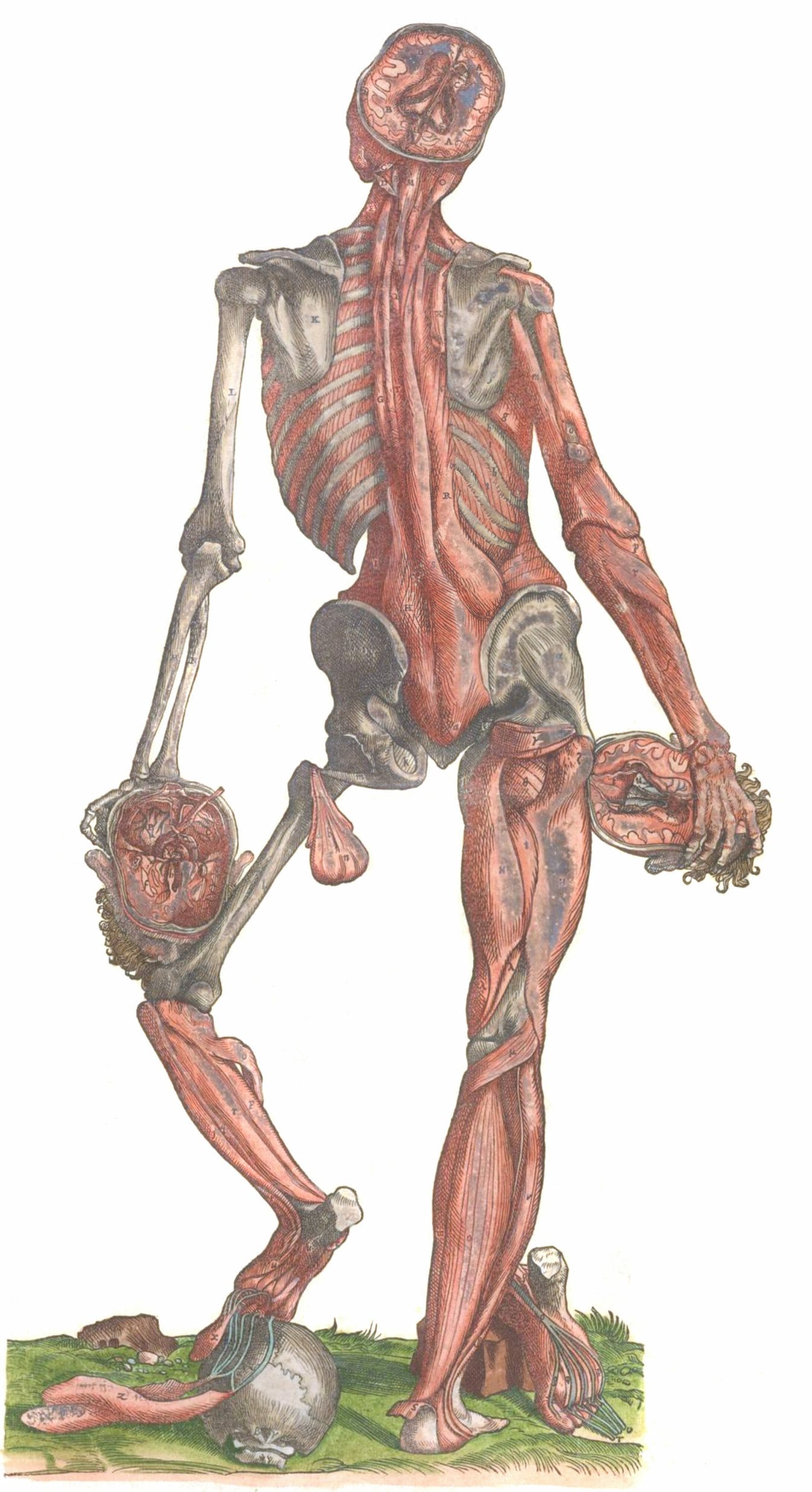
The anatomical images produced here suggest much more emotional and philosophical commentary than other vintage anatomy drawing at the time. It’s suggestive that this artist and scientist was interested in studying not just the physical elements of the body but wanted to have a holistic view of what makes us human, body and soul together.
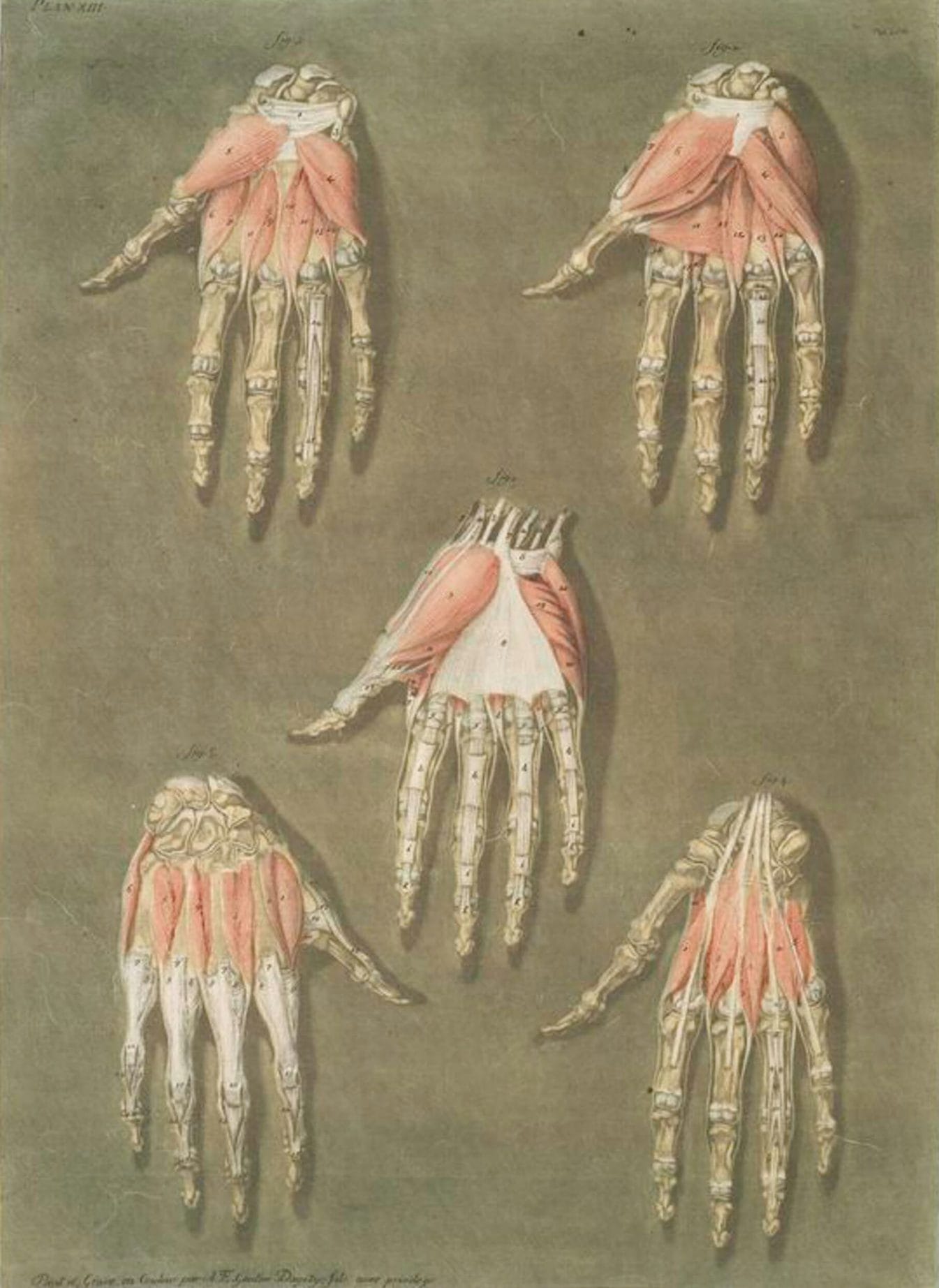
Illustration of the muscle structure of the face and hands
YIKES. Now’s we’re getting really creepy. These vintage medical paintings are by Arnauld Eloi Gautier-Dagoty, and they’re unsettling to say the least.
The way he’s shown the muscles and tendons on the bones of the hand in this illustration gives us the feeling that they’re about to start moving, despite being detached from a body. The numbered labeling brings us back into the scientific realm, and we can see how detailed and correct this drawing is.
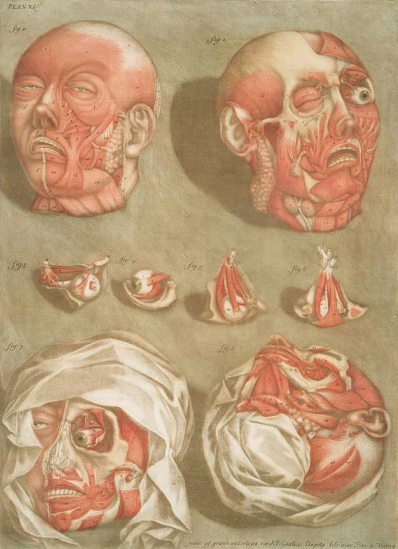
As for the severed head, this is the stuff of nightmares in picture form. Can you imagine what it would have been like to have the real thing sitting in front of you while you sketched away? The muscle structure is drawn here in perfect detail, and it shows where everything sits on the skull, but the things that gets me is the expression on the face. The downturned mouth and exposed, staring eyeball give this head a look of shock and fear.
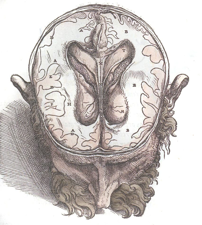
Anatomy murder and body snatching – the dark side of old anatomical illustrations
When we look at a medical diagram it’s easy to just see it as a scientific specimen. But illustrations like these in-detail pictures of the brain, where the face is drawn in such detail, that the individual characteristics of the person have been captured, remind us that most of the vintage medical illustrations we see would have been from life (or, death, really). It makes us think – who were these people that ended up as subjects in a laboratory? History has a few sources, some more chilling than others.
The Scientific Revolution was a period around 1550 – 1680. The uptake in scientific research and discovery was in many ways due to advances in book production, which mean that ideas could be shared much more widely than ever before. This spike though, mean that there was a demand for cadavers (dead bodies) for anatomical study, and the demand never dropped again, but only grew stronger. Unfortunately, some saw this as a lucrative opportunity and grave-robbing (body snatching) and murder became a more common way to provide scientists with specimens. There’s a particularly chilling account of two men in Edinburgh in the early 1900’s, who murdered a total of 16 people to provide bodies to anatomist Robert Knox, starting with tenants in their own boarding house. While this was obviously awful, the discovery of the murders led to new legislation around the provision of bodies for science.
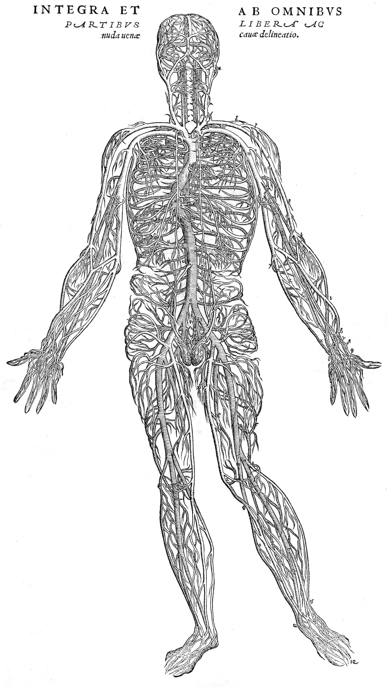
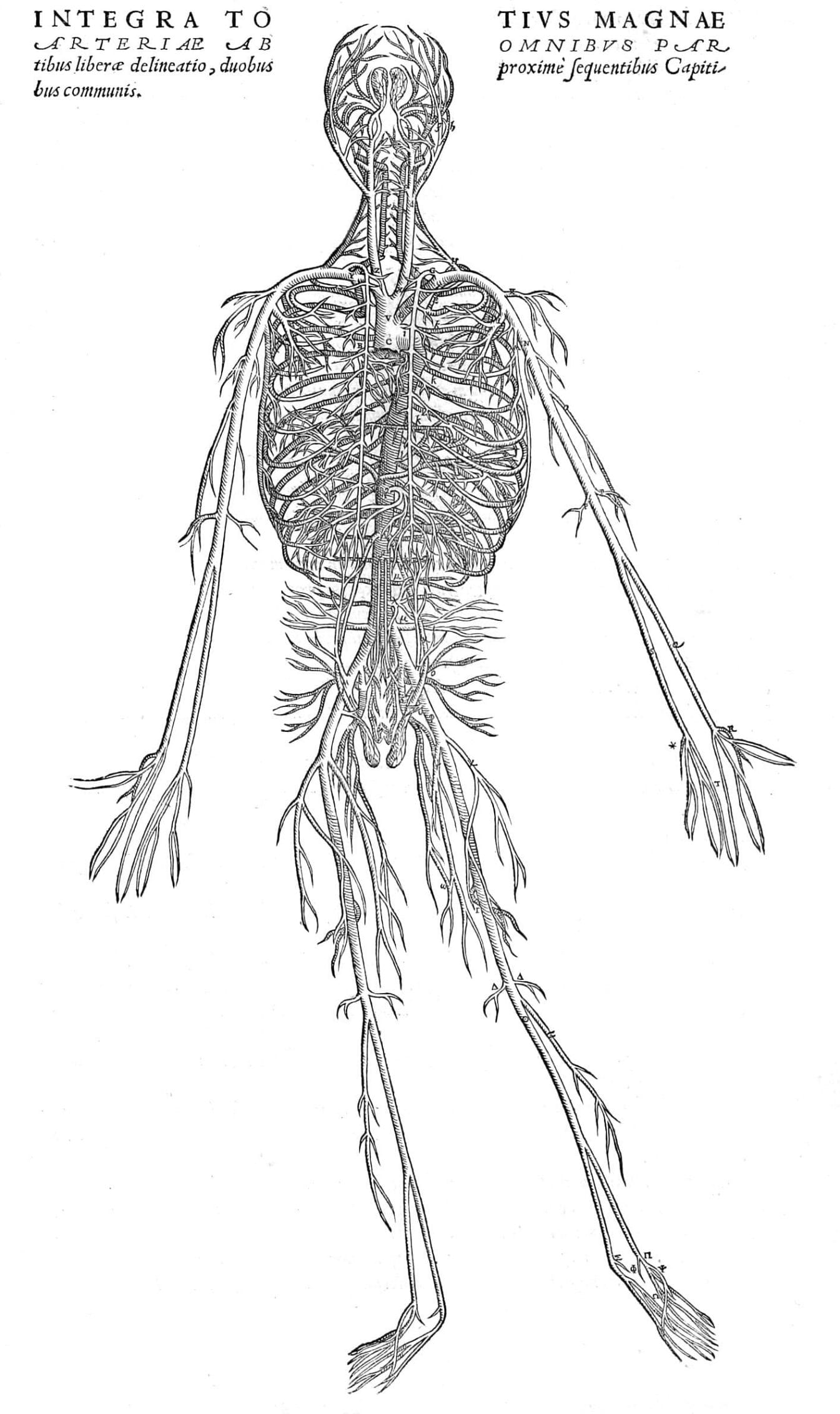
Antique medical illustration of the vascular system in human anatomy
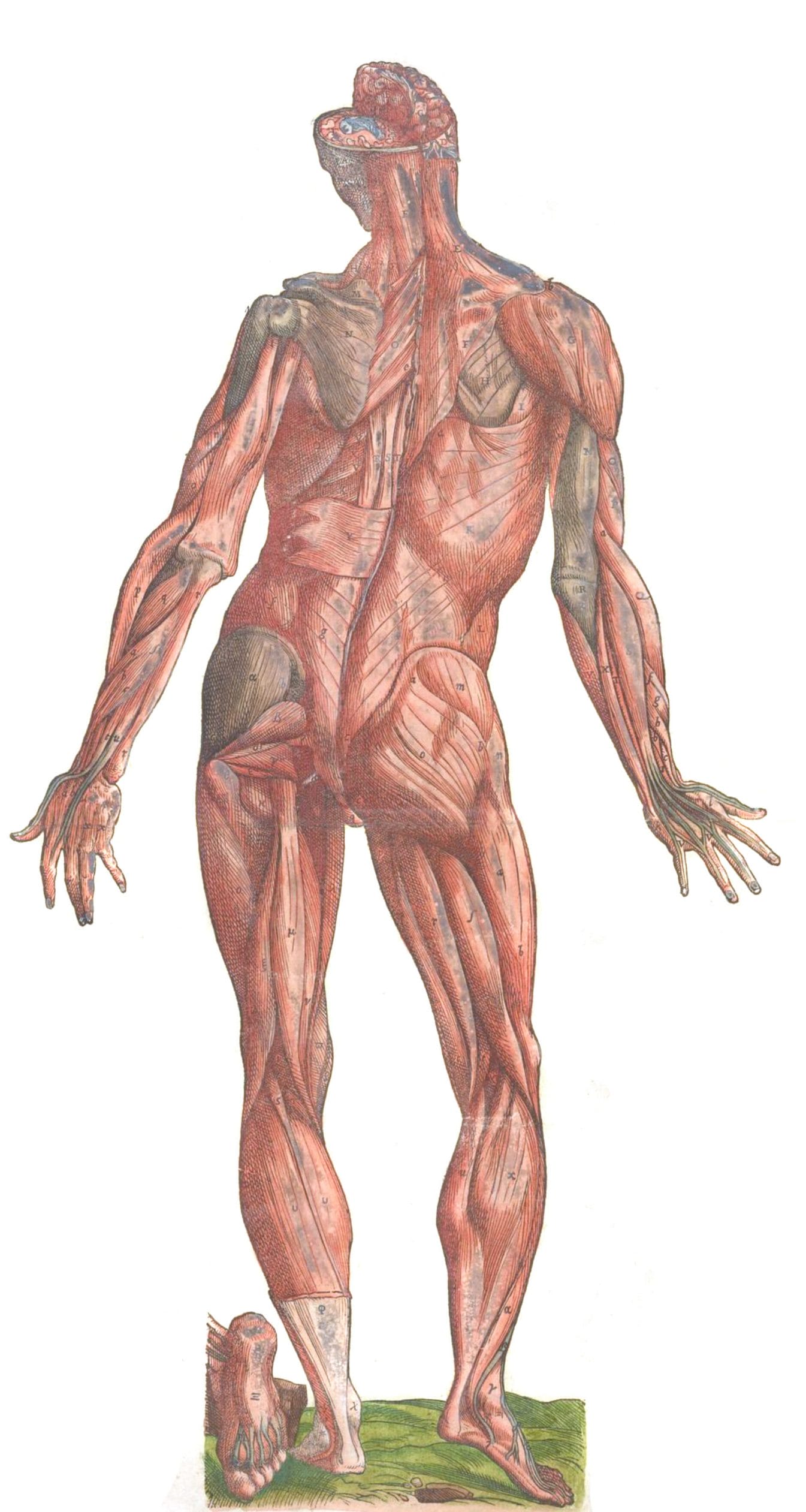
Another pair from Vesalius, this time with musical theatre hands! In this image, we have a very detailed depiction of the blood vessels in the body. I know, Vesalius produced extensive, important, and grotesquely beautiful work… but I forgot to mention before that he was known to use bodies that had been snatched for his work! That is, bodies that had been stolen from graves, often the graves of poorer marginalized people with little rights or support.
Want even more vintage anatomy illustrations? What you’ve seen here is just a tiny taste of what we have over at the full gallery. All the images you’ll find there are in the public domain, which means they’re free to use as you wish – for personal or commercial use!